

![]()
Principal Investigators: Sandra Shultz, Randy Schmitz, James Coppock, Sam Seyedin
Application Number: 62/755,817
UNCG Innovation: 18-0002
Category: Kinesiology
Description:
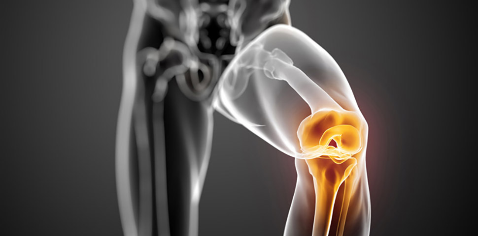
Instrumented knee ligament/laxity testing device would provide objective measures of knee joint rotational displacement in the frontal (VV, varus-valgus laxity) and transverse (IE, internal-external rotation knee laxity) plane and translational displacement in the sagittal (AP, anterior-posterior knee laxity) plane. AP laxity tests the isolate integrity of the anterior (ACL) and posterior (PCL) cruciate ligaments, VV laxity tests the isolated integrity of the medial and lateral collateral ligaments, and IE laxity tests the integrity of the both cruciate and collateral ligaments. This device would consist of a thigh stabilization element and a lower leg and foot stabilization element that would allow isolation of laxity testing in each plane of motion (while locked in the other two planes of motion) with a single positioning of the subject/patient. Precision sensors placed on the testing apparatus as well as on the limb could cancel out any movement artifact that would then accurately measure laxity in all three planes of motion (this part needs development of how it would actually work). Thee goal is that the device would: 1) allow a quick and easy measurement of tri-planar knee laxity, and 2) positioning instruction would not require an experienced examiner (thus removing as much tester error as possible), and 3) objective measures validated against gold standard (X-ray) could be documented.
The device with accompany software should be capable of measuring displacement at a fix load, and also constructing a load-displacement curve to quantify joint stiffness/compliance using accompanying software. The software would also calculate relaxation curves, and would allow input of subject height, weight and femur and tibia length so that laxity values can be normalized to body size (this is not a feature of current systems). The thigh and leg cradle would be equipped with a sliding caliper to accurately and efficiency record femur and tibia segment Length.
Immediate & Future Applications:
Can be used by physicians (family practice, orthopaedic), physician assistants, athletic trainers, physical therapists and other allied health professionals to access injury risk (pre-injury) screening and to examine joint integrity following injury (acute and chronic disease progression), following surgery and the recovery and rehabilitation process.
Specific applications include:
- Characterization of laxity profiles in healthy and diseased knees
- Monitor changes in knee laxity with maturation
- Screening to identify excessive knee joint laxity in the physically active to identify those at risk for ACL injury
- Monitoring disease progression in those with osteoarthritis and other relevant joint diseases
- Provide normative data across age (child to older adult) and sex for injury risk screening and satisfactory restoration of function post injury and surgery.
- Injury Diagnosis
Inventor info: Sandra Shultz
- https://kin.uncg.edu/about-us/our-faculty/sandra-shultz/
- https://www.researchgate.net/profile/Sandra_Shultz
- https://scholar.google.com/citations?user=ypg15uMAAAAJ&hl=en
- https://libres.uncg.edu/ir/uncg/clist.aspx?id=2284
- https://kin.uncg.edu/wp-content/uploads/2016/08/shultz_cv.pdf
- Complete List of Published Work (130 publications) Can be Accessed in MyBibliography:
Inventor Info: Randy Schmitz
- https://kin.uncg.edu/about-us/our-faculty/randy-schmitz/
- https://scholar.google.com/citations?user=mYq8KV4AAAAJ&hl=en
Inventor Info: Sam Seyedin
Inventor Info: James Coppock
Media:
- https://uncg.academia.edu/SandraShultz
- https://natajournals.org/doi/full/10.4085/1062-6050-501-17
- https://natajournals.com/doi/full/10.4085/1062-6050-45.5.445
- https://onlinelibrary.wiley.com/doi/full/10.1002/jor.22093
![]()